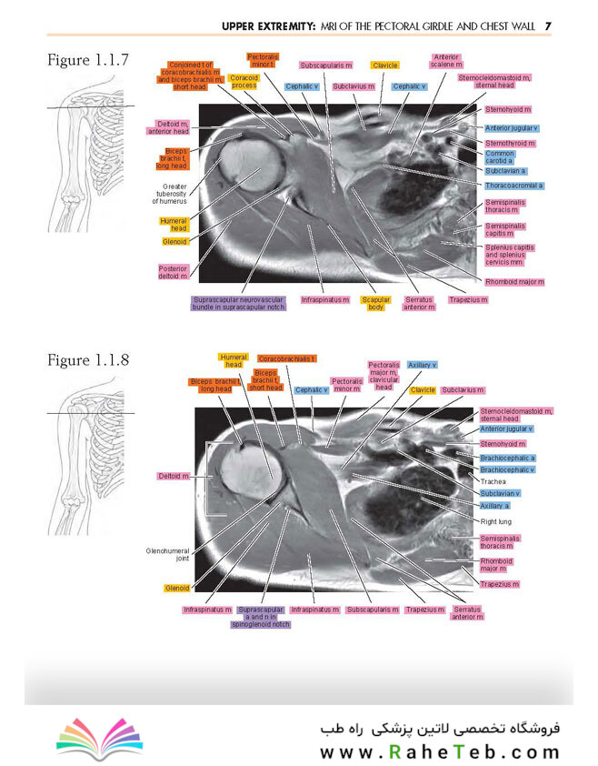
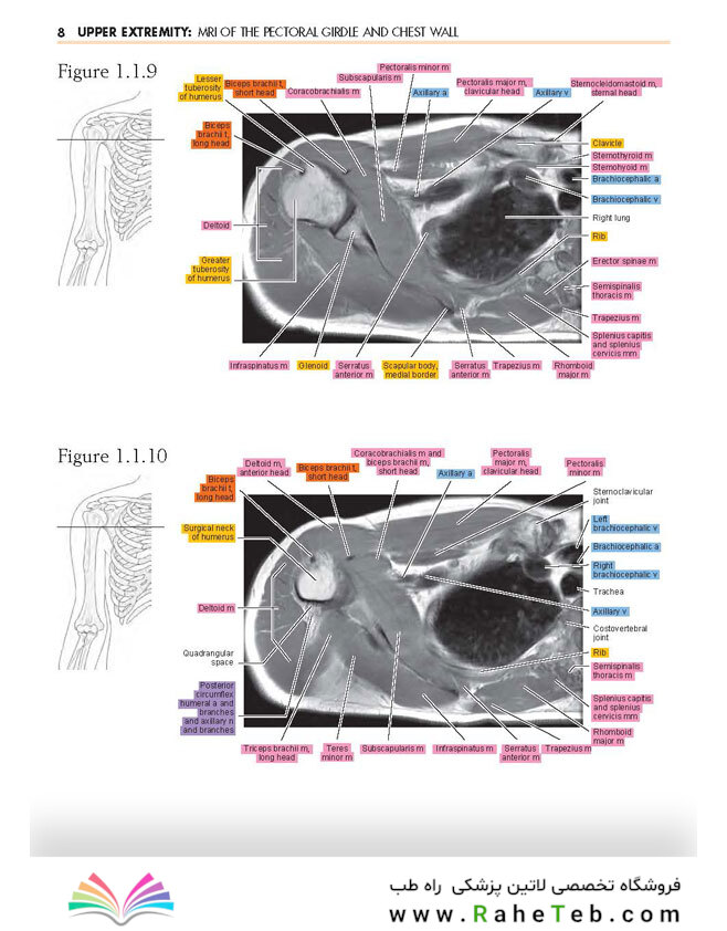
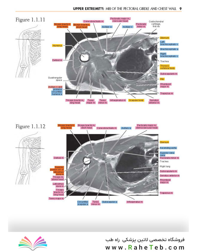
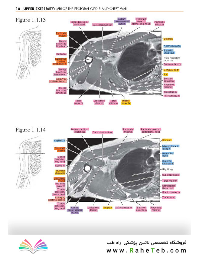
Sectional Anatomy by MRI and CT
کتاب آناتومی بخش MRI و CT
نسخه چهارم این مرجع کلاسیک که اکنون با پیشرفتهترین پیشرفتهای دیجیتال در دسترس است، حتی برای تمرین روزانه مرتبطتر و قابل دسترستر است. درک مطمئن آناتومی مقطعی برای تفسیر دقیق رادیولوژیک ضروری است و این اطلس دقیقاً اطلاعات مورد نیاز را در قالب مرجع عملی و سریع ارائه می دهد. کدگذاری رنگی جدید ساختارهای آناتومیک و قابلیتهای اسکرول و زوم جدید روی عکسها در نسخه کتاب الکترونیکی، این عنوان را به یک ابزار تشخیصی ضروری برای ساکنین و رادیولوژیستهای شاغل تبدیل کرده است.
Expert Consult نسخه کتاب الکترونیکی همراه با خرید. این تجربه پیشرفته کتاب الکترونیکی به شما امکان می دهد تمام متن، شکل ها، تصاویر و مراجع کتاب را در دستگاه های مختلف جستجو کنید.
برچسبهای رنگی برای اعصاب، عروق، ماهیچهها، تاندونهای استخوانی و رباطها شناسایی دقیق ساختارهای آناتومیک کلیدی را تسهیل میکنند.
قابلیتهای اسکرول و بزرگنمایی روی عکسها در نسخه کتاب الکترونیکی همراه، دسترسی آسانتر را در طول جلسات تفسیر و آموزش ساکنین زمان واقعی امکانپذیر میسازد.
MRIهایی که به دقت برچسب گذاری شده اند برای تمام اعضای بدن، و همچنین نمودارهای شماتیک و عبارات مختصر، ارتباط بین استخوان ها و بافت ها را روشن می کند.
سی تی اسکن برای قسمت های منتخب بدن، تجسم آناتومیک را افزایش می دهد.
بیش از 2300 تصویر پیشرفته را می توان در سه صفحه استاندارد مشاهده کرد: محوری، تاجی و ساژیتال.
Now available with state-of-the-art digital enhancements, the highly anticipated 4th edition of this classic reference is even more relevant and accessible for daily practice. A sure grasp of cross sectional anatomy is essential for accurate radiologic interpretation, and this atlas provides exactly the information needed in a practical, quick reference format. New color coding of anatomic structures and new scroll and zoom capabilities on photos in the eBook version make this title an essential diagnostic tool for both residents and practicing radiologists.
Expert Consult eBook version included with purchase. This enhanced eBook experience allows you to search all of the text, figures, images, and references from the book on a variety of devices.
Color-coded labels for nerves, vessels, muscles, bone tendons, and ligaments facilitate accurate identification of key anatomic structures.
Scroll and zoom capabilities on photos in the accompanying eBook version enable easier accessibility during interpretation sessions and real-time resident education.
Carefully labeled MRIs for all body parts, as well as schematic diagrams and concise statements, clarify correlations between bones and tissues.
CT scans for selected body parts enhance anatomic visualization.
More than 2,300 state-of-the-art images can be viewed in three standard planes: axial, coronal, and sagittal.
Product details
- Format Hardback | 624 pages
- Dimensions 222 x 281 x 30.48mm | 2,081.99g
- Publication date 31 Mar 2016
- Publisher Elsevier - Health Sciences Division
- Publication City/Country Philadelphia, United States
- Language English
- Edition Revised
- Edition statement 4th Revised edition
- Illustrations note Approx. 1165 illustrations (1165 in full color)
- ISBN10 0323394191
- ISBN13 9780323394192
- Bestsellers rank 1,322,889